Medial Lateral Gutter Loose Bodies
A The Anterolateral Gutter Is Demarcated Superiorly By The Inferior Download Scientific Diagram

Arthroscopic Treatment Of Capitellum Osteochondritis Dissecans With Micronized Allogeneic Cartilage Scaffold Arthroscopy Techniques

Inguinal Canal Anatomy And Physiology Textbook Medical Anatomy Pelvis Anatomy

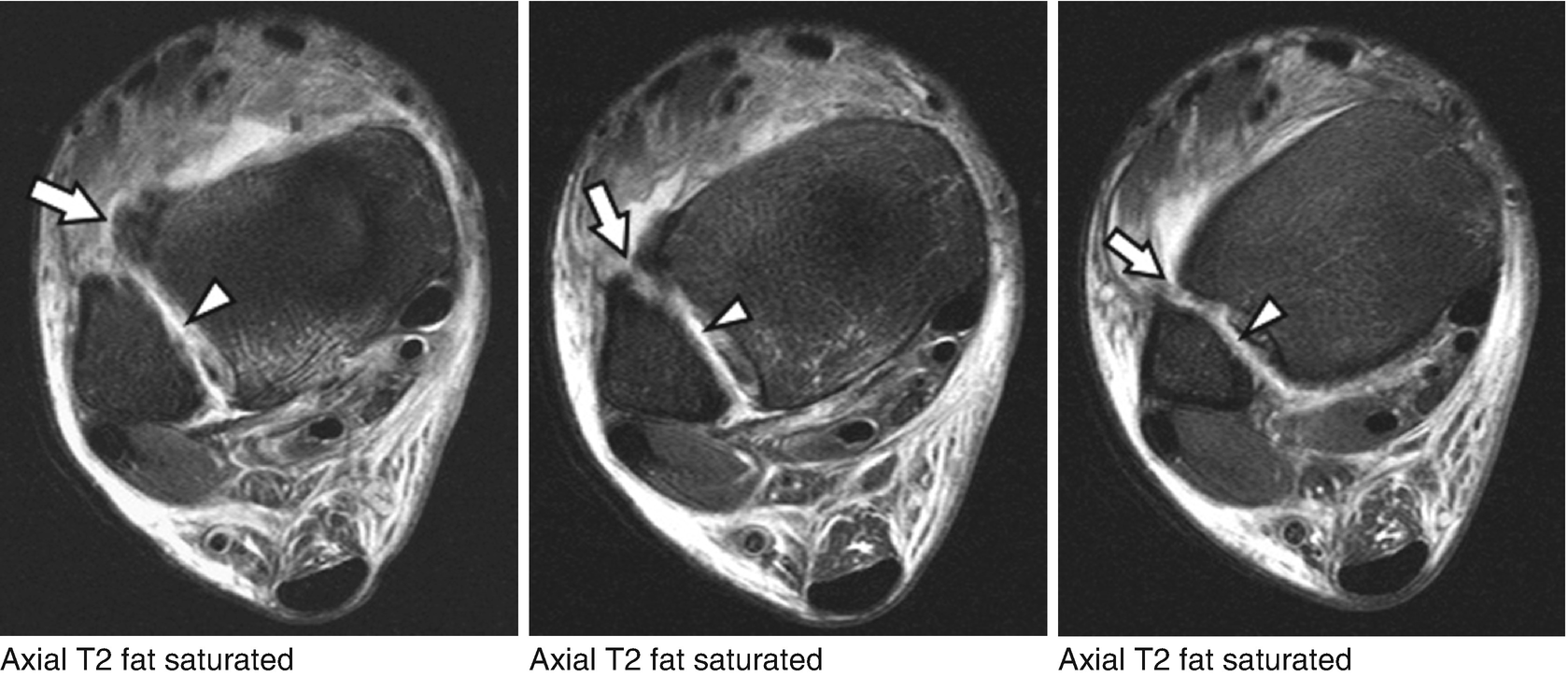
Osteochondral Loose Body In The Anterior Recess Of The Ankle Just Download Scientific Diagram

Wrist Ligament Injury I Had An Accident Tried To Break A Bolt Loose In A Situation Where A Whole Team Of Men Had Wrist Anatomy Sore Wrists Wrist Exercises

Abdominal Muscles Yoga For Osteoporosis Abs Workout Six Pack Abs Workout
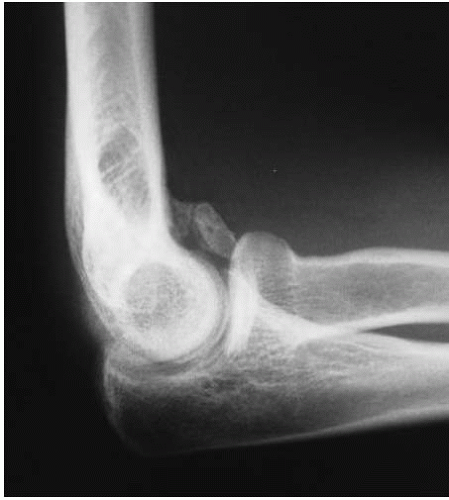
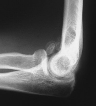
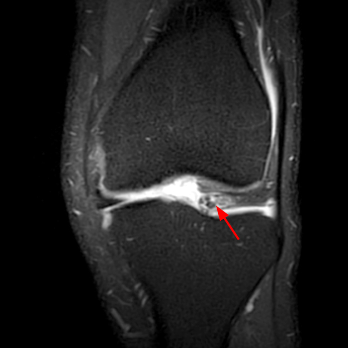
A 51 year old male who has developed a floating loose joint body around his left knee.
Medial lateral gutter loose bodies.

Anterior Triangle Of Neck Health Medicine And Anatomy Reference Pictures Anatomi Och Fysiologi Manniskokroppen Kropp

Intra Articular Loose Bodies Radiology Reference Article Radiopaedia Org

Lateral Right Ankle Radiograph With Evidence Of Calcified Loose Bodies Download Scientific Diagram
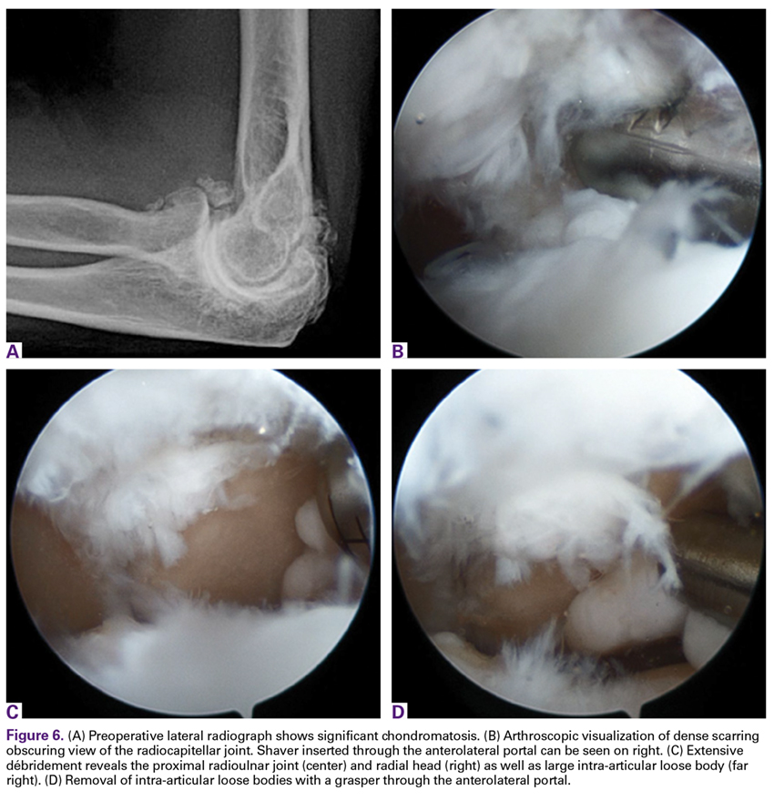
Diagnostic Elbow Arthroscopy And Loose Body Removal Musculoskeletal Key

Anteroposterior And Lateral Left Knee Radiographs Showing Typical Download Scientific Diagram
Ortho Blog Cmc Compendium

How To Handle Kinesiology Tape Infographic Kinesiology Taping Kinesiology Kinesio Taping

Aaos Bulletin April 2005

Image Picture21311493342704 Thumb For Term Side Of Card Human Anatomy Arteries Anatomy

Pin On Exercise For Back Muscles

Stretching The Psoas The So Az Is One Of The Most Inaccessible And Problematic Muscles In The Body Psoas Stretch Easy Workouts Stretches For Runners
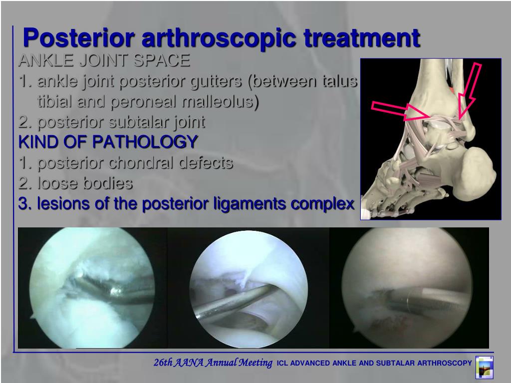
Ppt Advanced Ankle And Subtalar Arthroscopy Use Of Medial Portals In Ankle Arthroscopy Powerpoint Presentation Id 6639414

Stretching Exercises Encyclopedia Scoliosis Exercises Scoliosis Exercises Yoga Exercise

Ankle And Subtalar Arthroscopy Musculoskeletal Key

Protecting The Knee In Pigeon Pose Top Illustrates Engaging The Muscles On The Outside Of The Knee Bottom Show Pigeon Pose Piriformis Muscle Muscle Stretches

The True Hip Flexor Stretch Mike Reinold Psoasrelease Hip Flexor Stretch Hip Flexor Psoas Stretch
Diagnostic Elbow Arthroscopy And Loose Body Removal Musculoskeletal Key
Https Www Virtamed Com Files 6115 8335 0592 Vm Vba Arthros Brochure Us Print Version Pdf
Https Encrypted Tbn0 Gstatic Com Images Q Tbn 3aand9gcqpzkrh6xbudi6z4qi0tov1pc4aqggmkrf Vv4mnn0qa 7mic1g Usqp Cau
Synovial Plicae Of The Knee Radsource

Osteochondral Fracture And Patellar Instability Musculoskeletal Key

Pin On My Closet
5 Points On Stiff Elbow Mdedge Surgery

Degenerative Joint Disease Of The Ankle And Hindfoot Musculoskeletal Key
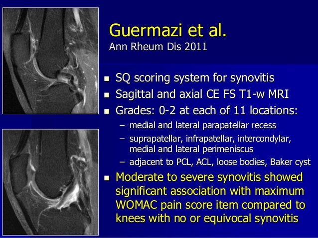
Imaging Of Synovitis In Oa

The Meniscal Grammar Signs Comma And Apostrophe Signs For Characterization Of A Displaced Fragment In The Meniscal Recess Sciencedirect

Management Of Talar Component Subsidence Foot And Ankle Clinics

Can Knee Braces Help You Recover From A Torn Meniscus Injury Mcdavid

Amazon Com Kohm Kp 700 Toenail Clippers For Thick Ingrown Nails Surgical Grade Stainless Steel 5 Long Inc Toe Nail Clippers Ingrown Nail Ingrown Toe Nail
Ankle Foot Springerlink

Fresh Osteochondral Allograft Transplantation For Uncontained Elongated Osteochondritis Dissecans Lesions Of The Medial Femoral Condyle Arthroscopy Techniques

Pin On Our Blogs

Ski Osteopati Og Fysioterapi Ligger Sentralt I Ski Sentrum Hos Oss Er Terapeutene Utdannet Bade Innen Osteopati Og Fysioterapi For Fysioterapi Utdannelse Ski

The Radiology Assistant Mri Examination

Elbow Arthroscopy In Trauma And Reconstruction Sciencedirect

Elbow Arthroscopy For Treatment Of Valgus Extension Overload Sciencedirect

Arthroscopic Treatment For Elbow Arthritis Osteophyte Removal Helps Regain Elbow Function

Arthroscopy Of The Foot And Ankle Musculoskeletal Key

Osteochondritis Dissecans Repair Musculoskeletal Key
Https Www Ajronline Org Doi Pdf 10 2214 Ajr 181 2 1810551

The Stiff Knee Clinical Gate
Gemc Injuries Of The Lower Extremity Knee Ankle And Foot Resident
Https Www Velocityhc Com Wp Content Uploads 2019 09 Coding Arthroscopic Knee Procedures Pdf
Https Encrypted Tbn0 Gstatic Com Images Q Tbn 3aand9gcqiyqyfrdh0endzhav7jxzuptotco8ktjbfenssc S756e0q5id Usqp Cau
Source : pinterest.com
